• English
  • العربية
  • 中文
  • Français
  • Русский
  • Español

You are here

How to Improve the Protection of Patients Undergoing Frequent Medical Imaging

,
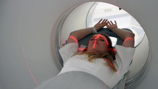
A patient undergoing PET/CT study

A patient undergoing a PET/CT scan. (Photo: L. Dojcanova/IAEA)

Progress made to reduce radiation-related risks – while maintaining the benefits – for patients who need frequent medical imaging was discussed at a virtual meeting held by the IAEA this week. Participants covered the impact and concrete actions needed to strengthen patient protection guidelines and technological solutions to monitor patient exposure history and took stock of global efforts to continuously enhance radiation protection of patients.

“Every day, millions of patients benefit from diagnostic imaging such as computed tomography (CT), X-rays and image-guided interventional procedures nuclear medicine procedures but with the increased  use of radiation imaging comes the concern about the associated increase of radiation exposure for patients,” said Peter Johnston, Director of the IAEA Radiation, Transport and Waste Safety Division. “It is critical to establish concrete measures to improve justification for such imaging and optimization of radiation protection for each patient undergoing such diagnosis and treatment.”

Over 4 billion diagnostic radiological and nuclear medicine procedures are performed globally each year. The benefits of these procedures far outweigh radiation risks when they are performed only as clinically justified, using the minimum necessary exposure to achieve the required diagnostic or treatment objective.

The radiation dose from a single imaging procedure is very low, ranging typically from 0.001 mSv to 20-25 mSv, depending on the type of the procedure. This is comparable to the natural background radiation exposure a person gets from a few days to a couple of years. “However, radiation risks may heighten when a patient undergoes a sequence of imaging procedures involving radiation exposure, especially if they are performed within short periods of time,” said Jenia Vassileva, an IAEA Radiation Protection Specialist.

Over 90 experts from 40 countries, 11 international organizations and professional bodies attended the meeting from 19 to 23 October. Participants included radiation protection experts, radiologists, nuclear medicine physicians, clinicians, medical physicists, radiation technologists, radiobiologists, epidemiologists, researchers, manufacturers and patient representatives.

Tracking radiation exposure of patients

Accurate and regular recording, reporting and analyzing of patient radiation doses in medical centres can help enhance dose management without the loss of diagnostic information. The tracked information from a patient’s previous examination and doses can contribute to preventing unnecessary exposures.

Highlighted at the meeting was  a IAEA  2009 project— Smart Card project —  that currently forms the foundation of a methodology for tracking exposure history. This project encouraged the industry to develop electronic systems and software solutions for exposure monitoring and tracking as more doses mean higher risks for patients.

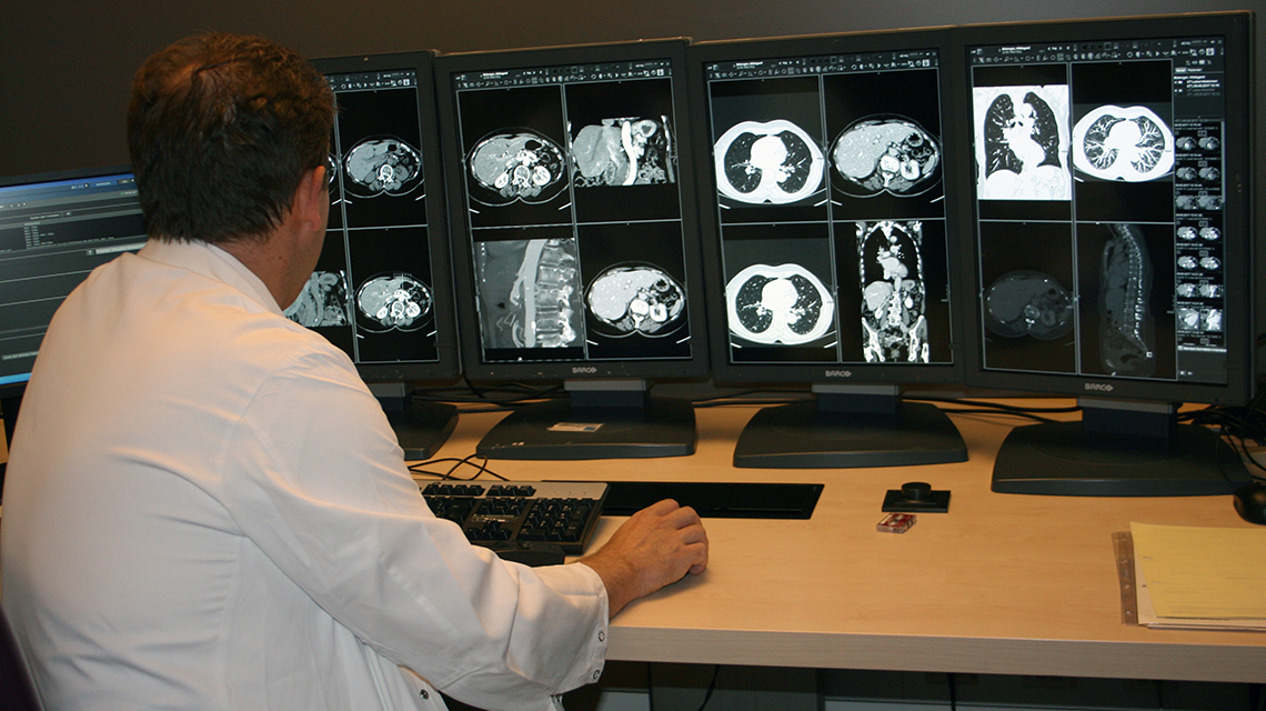
A radiologist reviewing CT scans of a patient. (Photo: L. Dojcanova/IAEA)

“As a result of the wider utilization of radiation exposure monitoring systems, data available demonstrates that the number of patients who accumulate effective dose of 100 mSv and higher in a few years from recurrent computed tomography procedures is greater than previously known and estimated to be one million patients globally per year,” said Madan M. Rehani, Director of Global Outreach for Radiation Protection at the Massachusetts General Hospital in the United States and Chair of this meeting.  “One out of five such patients is likely to be below 50 years of age where radiation effects are of higher concern, if patients have a longer life expectancy and therefore a higher chance for cancer to develop due to higher radiation exposure.”

Effective dose measured in millisieverts (mSv) is a quantity describing the effects of radiation exposure on organ and tissues and is the main quantity used in radiation protection. It indicates the probability of long-term radiation effects, primarily risk of developing cancer from exposure. Based on the current knowledge from the epidemiological studies, higher dose contributes to this increased probability.

Way Forward

There is a need for effective and enhanced guidance for patients with long-term illnesses and conditions that require frequent imaging, participants concluded. They agreed that radiation exposure tracking needs to be utilized broadly and integrated with other health care information systems for best results. Additionally, they emphasized the need to further develop imaging machines that use lower doses and standardised dose monitoring software tools for use worldwide.

But machines and better systems by themselves are not enough. It is the users, including physicians, medical physicists and technicians who are responsible for the optimal use of such advanced tools. It is therefore important that they receive appropriate training and the latest information on radiation risks, share knowledge and experience and communicate openly and transparently about benefits and risks with patients and caregivers.

Stay in touch

Newsletter