
Lower back pain is the sixth most common cause of global disability in the word and is often worsened by current conditions of life such as sedentariness, car journeys, desk work... When disk herniation, lumbar spinal stenosis or severe lumbar osteoarthritis is responsible for intense back pain, spine surgery is often performed. Despite high success rates, this very specific and precise surgery can lead to complications of which the most serious is an infection, called spondylodiscitis. Metallic instrumentation, often used in spine surgery, fosters the infectious risk. Spondylodiscitis causes major acute and chronic damages that can lead to disabling sequelae. Because this post-operative infection requires an intensive and targeted antibiotic treatment, early diagnosis is crucial and relies mainly on radiological examinations. However, spinal infection imaging is complex and there remains uncertainty about the imaging modality of choice. Conventional X-Ray provides poor information and abnormalities detected on CT are unspecific and delayed compared to the evolution of the infectious disease. Thanks to its high tissue contrast resolution, MRI is the radiological examination of reference. Unfortunately, metallic instrumentation generates artefacts that hamper the image quality and the diagnosis confidence.
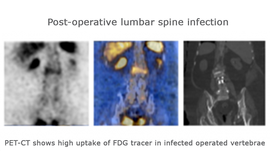
In order to determine the best imaging modality and to increase the performance of post-operative spinal infection imaging, two modern hybrid diagnostic techniques were tested and compared to MRI: 99mTc-UBI SPECT/CT and 18F-FDG PET/CT. Both combine scintigraphic images with radiological images (CT) to provide rich metabolic and anatomical information in the same exam. A specific infectious tracer, UBI labelled with 99mTc, was used for SPECT/CT and a glucose analogous largely uptake by spinal infection, FDG labelled with 18F, was used for PET/CT.
The general objective of is this CRP was to assess the role of multimodality imaging (MRI, 99mTc-UBI SPECT/CT, 18F-FDG PET/CT) for spine infection diagnosis after spinal surgery.
The specific Research Objectives was to:
- Determine the clinical value of MRI, 99mTc-UBI SPECT/CT and 18F-FDG PET/CT in the diagnosis of post-operative spondylodiscitis
- Determine if an imaging algorithm can be identified for the evaluation of post-operative spondylodiscitis
These 3 modalities were tested on patients suspected to have post-operative spinal infections and their results were compared to the disk biopsy or to the clinical evolution which determined if there was spinal infection.
At the end of the CRP, it appeared that 99mTc-UBI SPECT/CT was a poor diagnostic test and that MRI and 18F-FDG PET/CT were found equally as fairly useful tests. The 18F-FDG PET/CT offered a slight advantage due to its higher specificity and also because it provides a whole-body examination contrary to the MRI’s limited field of view to the spine.
For these reasons 18F-FDG PET/CT can be considered as a useful imaging tool for the diagnosis of spine infection. This modality could be classified in first position in the diagnostic imaging algorithm or be used as a complement to an MRI if its results are insufficient or equivocal.
Overall this CRP has increased the knowledge and strengthened the practice of the participating centres in the evaluation of post-operative spinal infection imaging and acknowledged the importance of diagnosing this severe disease early.
12 MSs countries participated in this CRP and 19 contracts were signed.

