
If you would like to learn more about the IAEA’s work, sign up for our weekly updates containing our most important news, multimedia and more.
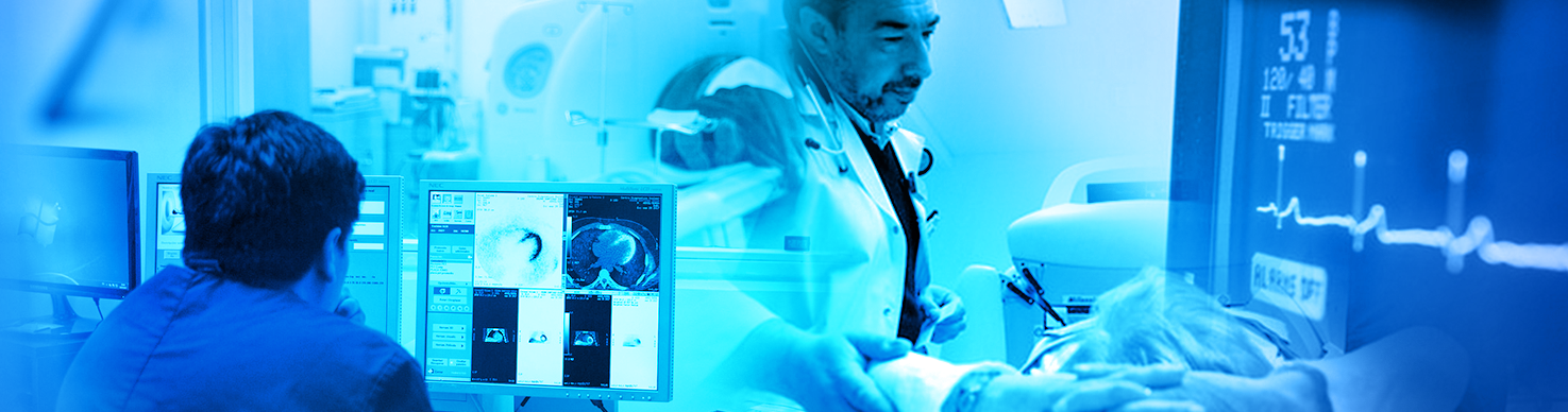
Dose and quality assessment of X-ray devices for interventional angiography and cardiology
Presenter: Prof. Nick Marshall, Prof. Hilde Bosmans, Michiel Dehairs
Date of broadcast: 16 October 2018, 3 pm CEST
About the webinar
X-ray systems for interventional angiography and cardiology are sophisticated and extremely flexible devices. Medical physics services are tasked with their commissioning and subsequent QC testing, with the aim of ensuring the safe use of these systems for both patient and personnel. These tests can also reveal aspects of system setup where the installed protocols can be optimized or better matched to clinical applications performed locally. QC testing therefore starts with an understanding of the interventional equipment, its architecture and intended use. What will be the common clinical applications? What is the specification of the X-ray tube, the X-ray focus options and the available spectral pre-filtration? How is the X-ray detector set up and operated?
Typical QC protocols therefore tackle the full system by assessing the different sub-components in turn, typically building up from the tube and generator. In this talk, we will step through the parts of a comprehensive QC test for an interventional imaging system. We begin, with a look at the applications set up on the system and the technical parameters that lie behind these imaging protocols. Most systems have an enormous number and variety of acquisition programs and it is a daunting task for the physicist to select the correct/relevant programs or modes for testing. It is generally impossible to test all applications available so some selection is required – it is important that tests assess frequently used imaging modes and protocols. This highlights one of the difficulties of testing such flexible devices: we want to make repeatable tests that can catch potential changes in system or component performance, yet we also want to assess how the system is set up for the common clinical applications.
We will look at means of assessing the automatic dose rate control (ADRC) system, including patient equivalent phantom selection, the implications for dosimeter type and use. We address the difficult but important topic of image quality evaluation using test objects. We cover detector specific methods and more generalized approaches that include the influence of the X-ray source and selected energy on system image quality.
Learning objectives
1. Understand the importance of quality assurance for ensuring the safe use of X-ray systems for interventional angiography and cardiology
2. Understand the influence of system specification and setup on optimization of clinical protocols
3. Learn the steps of a comprehensive QC test for an interventional imaging system
4. Appreciate the role of the medical physics expert as an important team member in optimization
About the presenters
Nicholas Marshall
Nicholas Marshall, PhD, was employed in the Radiation Safety Section of the Clinical Physics Group at Barts and The London NHS Trust from 2001 to 2009 to act as a Medical Physics Expert with a wide range of duties. These included managing both the routine QA testing and commissioning of all types of diagnostic radiology equipment and training of junior staff in all aspects of the QA of diagnostic radiology equipment.
From 2009 onwards, he worked in the KU Leuven (Belgium), first on the FP7 EU project breast-CT. In 2012 he joined the medical physics team of the University Hospitals Leuven and was appointed professor in 2014.
Prof Marshall’s domains of interest are the characterisation of detector technology for improved QA testing (especially in 2D mammography and breast tomosynthesis), interventional radiology, signal detectability studies and patient dose optimization. He is now project partner in the Horizon2020 project P3 STROKE, on which behalf present lecture on proper testing high end interventional and cardiological systems will be given.
Hilde Bosmans
Hilde Bosmans is professor in medical physics in radiology at the KU Leuven. Her medical physics activities include all modalities in x-ray imaging and more in particular also interventional radiology and cardiology. Next to clinical medical physics activities she also guides PhD students in medical physics, of which one student specifically focuses on improved quality and use of X ray dose in the cath lab. She was involved on several EC supported projects dealing with interventional radiology such as FP5 Dimond III, FP6 Sentinel and more recently Horizon2020 P3 Stroke, an EIT Health project. In this project, a new angio suit is being developed in which an x-ray tube and MRI system are combined into a single unit for stroke and other procedures. In the frame of this project, basic radiation protection measures have been reviewed.
Michiel Dehairs
Michiel Dehairs is a PhD student in medical physics in radiology at the KU Leuven. His field of research is interventional radiology and cardiology and more particular how the choice of X-ray factors influences the overall system efficiency. He is involved in the Horizon2020 P3 Stroke project, where he is working on radiation protection, X-ray image quality and system optimization for the Angio-MR system.